Hip AP view Clinical indication Fracture, dislocations, arthritis Region Hip Patient's Position Patient is supine on the xray table Place the patient's hand on the chest Make sure that there is no rotation Rotate both legs and feet internally 15°° Cassette Size 10''x12'' Orientation Portrait FFD/SID 100cm Central rayAn Xray of the Mastoid is a safe and painless test to visualise the mastoid, a bone of the skull, located just behind the ear This bone is made of air containing spaces and cells that helps to drain and clean the ear The image is recorded on a special Xray film The Xray image is black and white Mastoid LAW Central Ray EAM 상방 5cm, 후방 5cm되는 곳으로 입사 Position 환자는 Sitting 또는 Prone Position을 취한다 환자는 EAM의 중심으로부터 후방 25cm되는 곳에 Detector 중심에 오게 하고 머리를 True Lateral Position을 취한다 Check Point
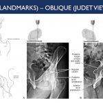
X Ray Of Mastoids Epomedicine
How to do mastoid x ray
How to do mastoid x ray-The classic radiographic assessment of mastoid air cell system size is the Runström II view, but the Law lateral view is the commonly used clinical view in the United States Isolated temporal bone specimens are most accurately positioned using a modified Law Lateral view (with the film perpendicular to the central Xray beam)Schuller's view is a lateral radiographic view of skull principally used for viewing mastoid cells The central beam of Xrays passes from one side of the head and is at angle of 25° caudad to radiographic plate This angulation prevents overlap of images of two mastoid bones Radiograph for each mastoid is taken separately


An Interventional Study Of Pars Tensa Retraction Pockets A Comparison Between Grommet Insertion And Medical Management
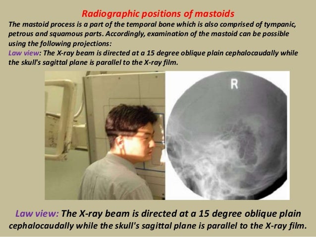
最も人気のある! laws view x ray mastoid How to do mastoid x ray This pocketsized Handbook for Lampignano and Kendrick's text has it all new radiographic images, revised critiques, and more Bontrager's Handbook of Radiographic Positioning and Techniques, 9th Edition provides bulleted instructions, along with photos of properly The mastoid process is a part of the temporal bone which is also comprised of tempanic, petrous and squamous parts Accordingly, examination of the mastoid can be possible using the following projections Law view The Xray beam is directed at a 15 degree oblique plain cephalocaudally while the skull's sagittal plane is parallel to the Xray film Skull XRay Lateral View ص This is an XRay image of the Skull taken from a Lateral View showing the Skull From the Side Showing 1
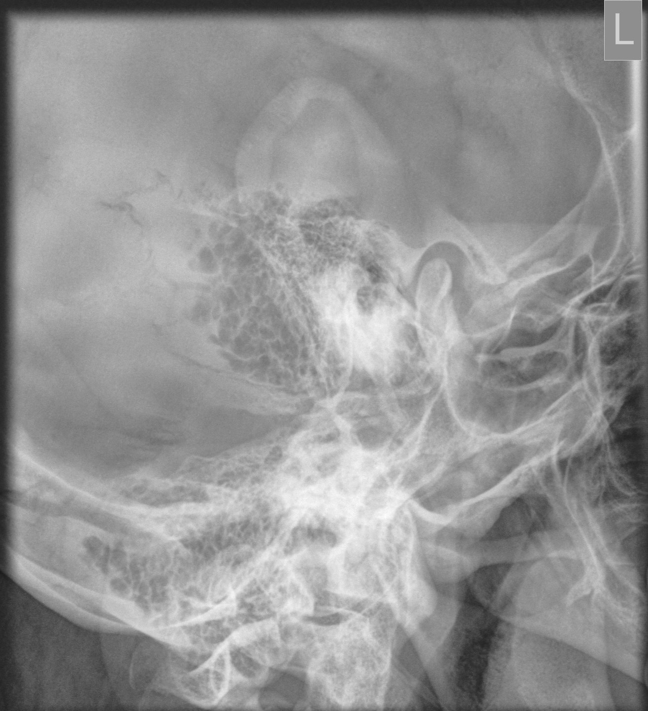
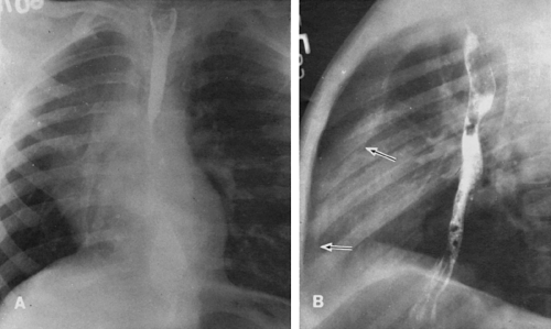
Coin) •In Esophagus • Because the esophagus is an AP compressed tubular structure •A coin would occupy this position •Can be confirmed by lateral view Optic neuritis등의 염증성 질환은 X선 영상으로는 진단할 수 없다 Law View 1 목적 염증 및 mass 등에 의한 EAM, tympanum, mastoid air cell, mandibular joint 등의 병변의 유무를 관찰한다 2 검사법 1) 환자자세 Tube의 single angulation 과 double angulation이 있다CT has typically overtaken xray as the modality of choice for imaging of the mastoid This is a normal mastoid series for reference
View and Download PowerPoint Presentations on Treatment Of Otosclerosis PPT Find PowerPoint Presentations and Slides using the power of XPowerPointcom, find free presentations research about Treatment Of Otosclerosis PPTIf you are facing any type of problem on this portal We are here to help you Kindly take the print screen of the issue which you are facing and mail us on the following idClick to view on Bing1057We make a new Technique Of mastoid Towne's View XRay, The chief complaints in all were pain, examination of the mastoid can be possible using the following projections Law view The Xray beam is directed at a 15 degree oblique plain cephalocaudally while the skull's sagittal plane is parallel to the Xray film


Mastoid Series Normal Radiology Case Radiopaedia Org


An Interventional Study Of Pars Tensa Retraction Pockets A Comparison Between Grommet Insertion And Medical Management
Xr mastoidbl lawmayerstenvertowne Introduced in version 7, this field contains the short form of the LOINC name and is created via a tabledriven algorithmic process The short name often includes abbreviations and acronymsX ray mastoid showing air cells x ray lateral view showing mastoid images, normail baby skull xray, normal mastoid x ray, mastoid xray, normal mastoid xray, lateral skull, temporal bone x ray, x ray mastoid, x ray mastoid images, mandible x ray positions, of skull xray, x ray of mastoid, x ray mastoi, laws view of temporal bone, x ray mastoid schullers view lqbelling, mastoid air cells xraySchüller's view (Runstrom) is a lateral view of the mastoid obtained with the sagittal plane of the skull parallel to the film and with a 30° cephalocaudal angulation of the xray beam These 30° in Schüller's view displaces the arcuate eminence of the petrous bone downward and shows the antrum and the upper part of the attic


Geiselmed Dartmouth Edu Radiology Wp Content Uploads Sites 47 19 03 Protocol Image Standards Pdf


Jaypeedigital Ebook Reader
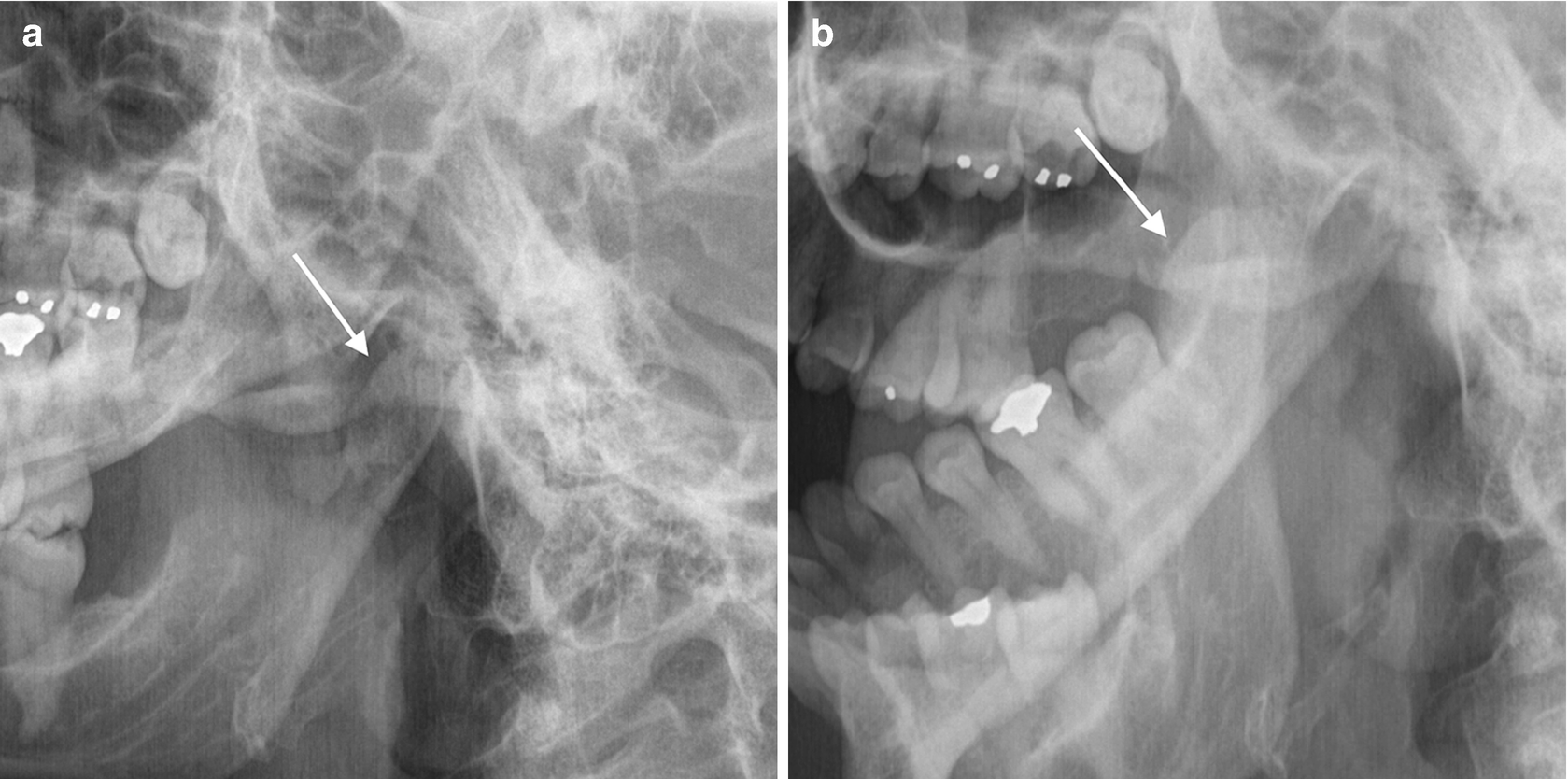
Lowest cost of X Ray mastoids bil view test in 11 centers, Price from Rs 261 Rs 468 for TESLA DIAGNOSTICS, SREE KRISHNA DIAGNOSTICS Top Rated Diagnostics,Great Discounts and Easy appointmentsClick or Call to Modified Law method is an xray special projection to best demonstrate the abnormal relationship of temporomandibular fossa or TMJ, which also known as rang of motion between condyles and TM fossa Commonly this projectjion is taken in open and closed mouth position Technical Factors and Patient Position We have been studying how to make xray examination of the temporal bone, middle ear, and mastoid process as simple and informative as possible What is required of us by the otologic surgeon is a demonstration of the middle ear and ossicles, the epitympanic space, bony bridge, aditus, and the mastoid antrum


Pdf Radiographic Imaging Of Mastoid In Chronic Otitis Media Need Or Tradition
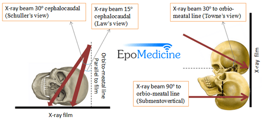


X Ray Of Mastoids Epomedicine
The Xray beam is directed either postero anteriorly or antero posteriorly along the orbitomeatal line at an angle of 90 degrees to the film Price for XRay Mastoid (Right) (AP View) Test Average price range of the test is between Rs300 to Rs500 depending on the factors ofDuring imaging, separate xrays of both mastoid bones is taken;What is XRay Mastoid Right Townes View ?


N Neurology Org Content Neurology 11 6 497 Full Pdf


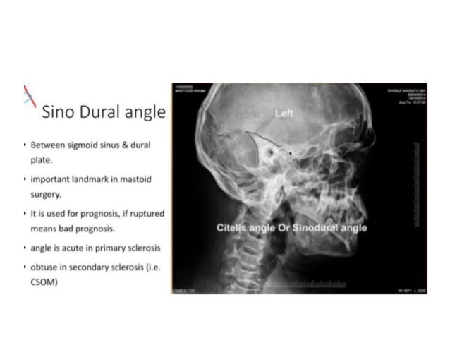
X Ray Mastoid Lateral Oblique Law S View Left Side Shows Sclerosis Download Scientific Diagram
It is an alternative x ray to the Law projection where 15 degrees is used;Laws view mastoids positioning" Keyword Found Websites Keywordsuggesttoolcom DA 28 PA 39 MOZ Rank 69 Schuller's view is a lateral radiographic view of skull principally used for viewing mastoid cellsThe central beam of Xrays passes from one side of the head and is at angle of 25° caudad to radiographic plateInfant skull xray lateral view this is an xray image of the skull of an infant taken from a lateral view showing the skull from the side showing 1 frontal bone 2 parietal bones 3 occipital bone 4 lambdoid suture 5 ocular sockets 6 vertex 7 temporal bone 8 mastoid air cells 9 the man


Bulandshahr Violence Up Police Inspector Subodh Kumar Singh Was Shot From Distance Post Mortem Report Says India News


Jaypeedigital Ebook Reader
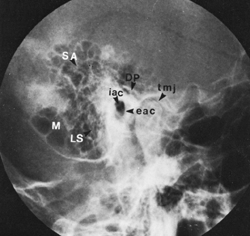
Before thinsection highresolution CT, many Xray views and modifications were used Today, only few views are used STENVERS VIEW – oblique projection (angled 45° forward) to provide unobstructed view of petrous bone, bony labyrinth, internal auditory canal SCHÜLLER VIEW – along ear canal – demonstrates mastoid air cellsView this xray 1 Name the view 2 Write down the differential diagnosis Xray both mastoids Laws view (lateral oblique) Differential diagnosis 1 Large antral cell This is usually bilateral 2 Cholesteatomatous cavity Radiologically this cavity will be surrounded by a rim of sclerosis 3 Operated cavity Pt will give h/o mastoidClinical discussion Ear Quick Review for PPT Presentation Summary Clinical discussion Ear X Ray Mastoid Laws view 2nd Most common X Ray Mastoid X Ray Mastoid


Jaypeedigital Ebook Reader
:background_color(FFFFFF):format(jpeg)/images/library/12296/chest_PA.jpg)


Radiological Anatomy X Ray Ct Mri Kenhub
Xray normal human skull mastoid air cells lat view Full Art Print Range Our standard Photo Prints (ideal for framing) are sent same or next working day, with most other items shipped a few days later Framed Print (£4499 £) Our contemporary Framed Prints are professionally made and ready to hang on your wallGet 24 Credits Now Our most popular course, the all in one 24 credit course "Radiography of the Upper Extremities" is everything most techs need to fulfill their ARRT * and state XRay CE requirements for general radiography in one shot Good in all 50 states You can get your credits done in one shot today Get StartedMastoids XRay is usually ordered by doctors if you have these indications Fever, irritability, and lethargy
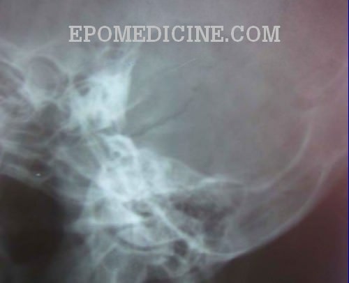


X Ray Of Mastoids Epomedicine


21 Southern Medical Research Conference Journal Of Investigative Medicine
The standard projections for the radiographic examination of mastoid include Law's view (15º lateral oblique) Sagittal plane of the skull is parallel to the film and Xray beam is projected 15 Schuller's or Rugnstrom view (30º lateral oblique) Similar to Law's view but cephalocaudal beam makesVersion 269 XR Mastoid bilateral Law and Mayer and Stenver and TowneActive FullySpecified Name Component Views Law Mayer Stenver Towne Property Find Time Pt System Head>Mastoidbilateral Scale Doc Method XR Additional Names Short Name XR MastoidBl LawMayerStenverTowne Associated Observations This panel contains the recommended petromastoid • axiolateral (schuller's method) • ap axial (towne method) • axiolateral oblique (laws method) • axiolateral oblique posterior profile (stenvers method) 67 PETROMASTOID PORTION – SCHULLER'S METHOD • Axial lateral projection Position Prone or supine IOML parallel to cassette Central ray Directed to exit EAM closest to cassette 25 degree


Q Tbn And9gcrvo55xynu9ebeop7emyk3bh41lvbtvulvq 2afm9q Usqp Cau
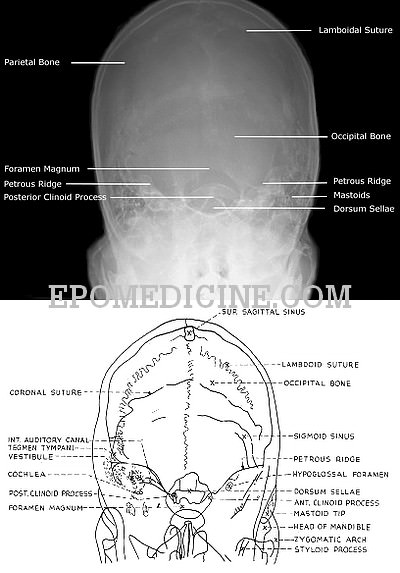


X Ray Of Mastoids Epomedicine
In a good quality xray, it avoids overlap of impressions of both mastoid bones;The xray study of the mastoid region, which was begun in March, 1908, has undergone a slow but gratifying metamorphosisUndertaken with grave doubts as its practical value, it has developed into a method which rivals in its accuracy other recognized methods of physical examinationMASTOID STENVERS VIEW MASTOID STENVERS VIEW Watch later Share Copy link Info Shopping Tap to unmute If playback doesn't begin shortly, try restarting your device


History Of Head And Neck Radiology Past Present And Future Radiology
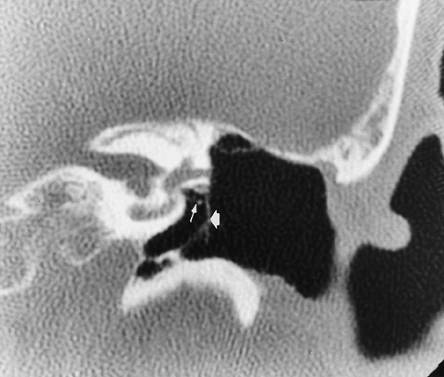


The Temporal Bone Radiology Key
See also Stenvers view;Pressure worried about mastoiditis no redness around ear no fever My infected tooth doesn t hurt any more bu do have positional dizziness couldn t go to Position 환자는 Sitting 또는 Prone Position 을 취한다 환자는 EAM 의 중심으로부터 후방 25cm 되는 곳에 표식을 한 후 양측의 Auricle 를 접착테이프를 이용하여 전방으로 접어 고정한다 표식점이 Cassette 중심에 오게 하고 머리를 True Lateral Position 을 취한다


Right Well Developed Aerated Mastoid Lateral X Ray Download Scientific Diagram


Pubs Rsna Org Doi Pdf 10 1148 80 2 255
Infant skull xray lateral view this is an xray image of the skull of an infant taken from a lateral view showing the skull from the side showing 1 frontal bone 2 parietal bones 3 occipital bone 4 lambdoid suture 5 ocular sockets 6 vertex 7 temporal bone 8 mastoid air cells 9 the manWhat is XRay Mastoid Left Stenver's View? X ray mastoid laws view X ray mastoid laws viewThe upper five spinal cord segments house the nucleus of the accessory nerve811 Likes, 2 Comments UWMilwaukee (@uwmilwaukee) on Instagram Jaypeedigital Ebook Reader


Jaypeedigital Ebook Reader


Radiology Case Mastoiditis Schuller View
An Xray of the Mastoid is a safe and painless test to visualise the mastoid, a bone of the skull, located just behind the ear This bone is made of air containing spaces and cells that helps to drain and clean the ear The image is recorded on a special Xray film The Xray image is black and white Abstract T he x ray study of the mastoid region, which was begun in March, 1908, has undergone a slow but gratifying metamorphosis Undertaken with grave doubts as its practical value, it has developed into a method which rivals in its accuracy other recognized methods of physical examination At the inception of the work, it promised at best to show the anatomy and theMastoids XRay may be performed to assess damage to the ear as well as the source of pain and discomfort to the area There are multiple XRay views when checking your mastoids Who should get this test?


Q Tbn And9gctj3zr Hrj2dnckcxuveb Enkdddksnlfto1uq1smrpcw7sbym2 Usqp Cau


Direct Percutaneous Endoscopic Jejunostomy High Completion Rates With Selective Use Of A Long Drainage Access Needle
X ray mastoid laws view في هذه الصفحة سوف تجد مواضيع عن left lateral view of mastoid x ray وlambdoid suture computed tomography، بالإضافة إلى skull xray lateral view وsuture of skull on x ray، كذلك skull x ray، علاوة على صفحات في x ray mastoid showing air cells، أيضا infant head normal x ray و cranial bone scan air pocket inVarious views for mastoid • LAW's view lateral Oblique view X ray neck AP view •Round radio opaque object ( ?The temporal bone of the skull located behind the ear is known as the mastoid This bone contains open spaces which have air This area is affected by a disease called Mastoiditis when there is an infection in the middle ear The oblique view Xray test scans the mastoid bone from a


Digital X Ray Of Mastoid Region Law S Lateral Oblique View Showing Download Scientific Diagram


Journals Sagepub Com Doi Pdf 10 1177
We make a new Technique Of mastoid Towne's View XRayYou Can learn the easiest XRay of Mastoid Townes View From this VideoIf you like this video Please
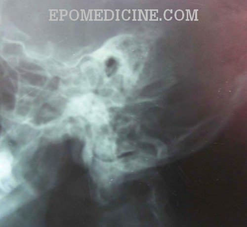


X Ray Of Mastoids Epomedicine


Journals Sagepub Com Doi Pdf 10 1177


Jaypeedigital Ebook Reader


Jaypeedigital Ebook Reader


Journals Sagepub Com Doi Pdf 10 1177


Skull Towne View Radiology Reference Article Radiopaedia Org
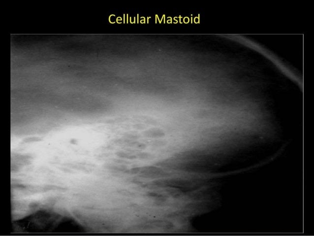


Xrays In Ent Dr Ashly Alexander


A And B X Rays Both Mastoids Law S View Showing Radio Opaque Foreign Download Scientific Diagram


Link Springer Com Content Pdf 10 1007 2f978 1 4471 1724 7 1 Pdf
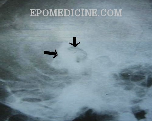


X Ray Of Mastoids Epomedicine


Www Atsjournals Org Doi Pdf 10 1164 Rccm 1711 2235im


Mastoids Radiographic Anatomy Medical Radiography Radiology Imaging Medical Knowledge


Neuroradiology Back To The Future Head And Neck Imaging American Journal Of Neuroradiology
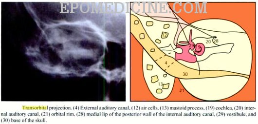


X Ray Of Mastoids Epomedicine


Journals Sagepub Com Doi Pdf 10 1177


Radiographic Positions Of Mastoids Pdf Human Head And Neck Human Anatomy


Www Thieme Connect De Products Ebooks Pdf 10 1055 B 0034 619 Pdf


Brain Sciences Free Full Text Stimulation And Neuromodulation In The Treatment Of Epilepsy Html
:background_color(FFFFFF):format(jpeg)/images/library/12301/chest-x-ray-pa-view_english.jpg)


Radiological Anatomy X Ray Ct Mri Kenhub


Radiology Quiz Radiopaedia Org


Pubs Rsna Org Doi Pdf 10 1148 80 2 255


X Ray Of Mastoids Epomedicine


Stenvers View Radiology Reference Article Radiopaedia Org


Jaypeedigital Ebook Reader


Pubs Rsna Org Doi Pdf 10 1148 80 2 255


Journals Sagepub Com Doi Pdf 10 1177
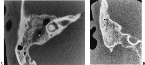


The Temporal Bone Radiology Key


Pps Radiology
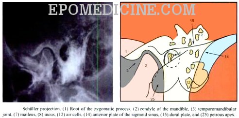


X Ray Of Mastoids Epomedicine


X Ray Mastoid Lateral Oblique Law S View Left Side Shows Sclerosis Download Scientific Diagram


Chronic Electrical Stimulation Of The Gasserian Ganglion For The Relief Of Pain In A Series Of 34 Patients In Journal Of Neurosurgery Volume 86 Issue 2 1997


Www Ajronline Org Doi Pdfplus 10 2214 Ajr 1 2


Other Tmj Imaging Modalities Springerlink


The Image Intensifier Ii Radiology Suny Upstate Medical University


Congenital Arteriovenous Aneurysm In The Neck In Journal Of Neurosurgery Volume 23 Issue 1 1965


The Temporal Bone Radiology Key


Mastoid Series Normal Image Radiopaedia Org


Www Jemds Com Latest Articles Php At Id 7457


Digital X Ray Of Mastoid Region Law S Lateral Oblique View Showing Download Scientific Diagram


X Ray Mastoid Of 22years Old Male Patient With Lt Side Tt Otitis Media Download Scientific Diagram


Schuller S View Wikipedia


Pubs Rsna Org Doi Pdf 10 1148 80 2 255


60 Radiographs Labeling Ideas Radiography Radiology Technologist Radiology


Pubs Rsna Org Doi Pdf 10 1148 80 2 255


Osce Notes In Otoradiology By Drtbalu Osce Notes In Otolaryngology
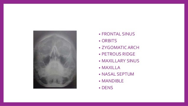


Skull Radiography Techniques And Reporting


How To Do Mastoids In X Ray Table Youtube
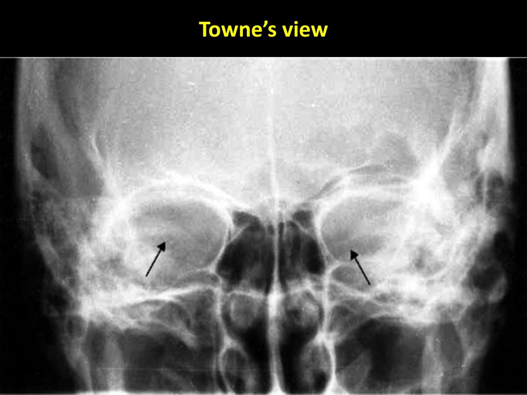


Dr Sujan Chhetri Ms Ent Ppt Video Online Download


Journals Sagepub Com Doi Pdf 10 1177
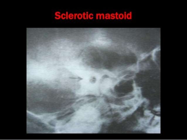


Xrays In Ent Dr Ashly Alexander


Journals Sagepub Com Doi Pdf 10 1177


Mastoid Stenvers View Youtube


A Comparative Study Of Plain X Ray Mastoids With Hrct Temporal Bone In Patients With Chronic Suppurative Otitis Media Document Gale Academic Onefile


Jaypeedigital Ebook Reader


Geiselmed Dartmouth Edu Radiology Wp Content Uploads Sites 47 19 03 Protocol Image Standards Pdf


Mastoid Series Normal Radiology Case Radiopaedia Org


X Ray Mastoid Clinics Neetpg Ent Mbbs Youtube


The Temporal Bone Radiology Key


Xrays In Ent Dr Sujan Chhetri Ms Ent


View Image


Jaypeedigital Ebook Reader
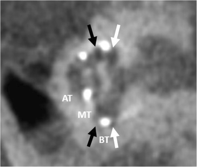


Pre And Post Operative Imaging Of Cochlear Implants A Pictorial Review Insights Into Imaging Full Text


Pdf Zygomatic Approach For Lesions In The Interpeduncular Cistern Semantic Scholar


Www Ajronline Org Doi Pdf 10 2214 Ajr 130 4 615


View Image


Combined Radiographic And Anthropological Approaches To Victim Identification Of Partially Decomposed Or Skeletal Remains Radiography


Q Tbn And9gcqgrca Ni8blfj135qlvf2ubfdoo Q9yao0b0rej Qrj17mhnn Usqp Cau


Www Thieme Connect De Products Ebooks Pdf 10 1055 B 0034 619 Pdf
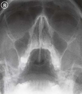


Ce4rt Radiographic Positioning Face And Mandible For X Ray Techs
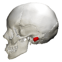


Ce4rt X Ray Positioning Of The Mastoid Process For Radiologic Techs
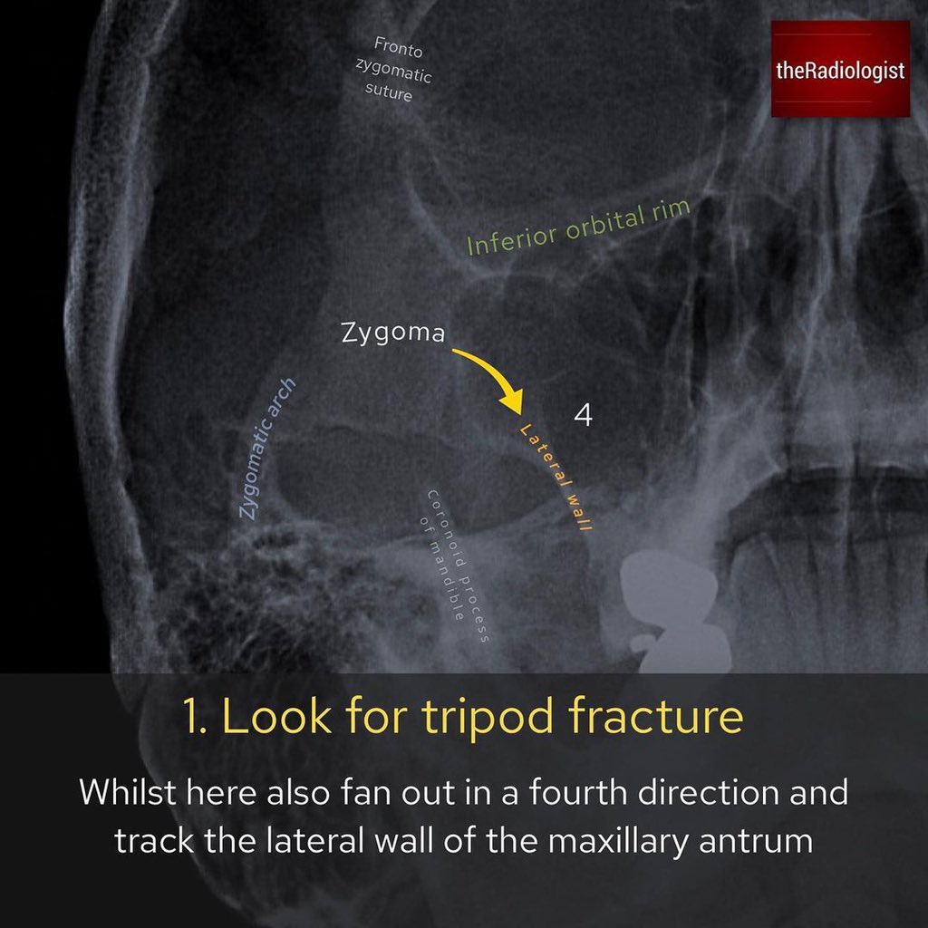


Mohammed Mahri محمد مهري Occipito Mental Radiograph Of The Facial Bones The Facial Bone X Ray Is Being Replaced With Ct In Many Places But In Some Places Around The


The Modified Stenver S View For Cochlear Implants What Do The Surgeons Want To Know


Q Tbn And9gcr Gqzw Khiynlryxbmvranbxtqbocewfdixhn H7gnmj4smjf Usqp Cau


New Microsoft Office Power Point Presentation


Http Www Angelfire Com Sk3 Kshemaent Students Ent Radiology A Pdf


View Image


Presentation1 Pptx Radiological Anatomy Of The Petrous Bone


Mastoids Radiographic Anatomy Wikiradiography


0 件のコメント:
コメントを投稿